English report
AS-HOPE 事業報告
事業番号:AS-24-008
To do my research and to present a paper at the international symposium on "Diversity and Evolution of Asian Primates"
報告者:Pomchote Porrawee
期間:2012/8/15 - 2012/9/27
1. To study age change of bone density using peripheral quantitative computed tomography (pQCT) and bone disorders such as osteophytosis and osteoarthritis using radiographic technique in live long-tailed macaques (M. fascicularis), which are non-seasonal breeders, reared at the Primate Research Unit, Department of Biology, Faculty of Science, Chulalongkorn University. I will determine whether there is a relationship between reproductive seasonality and aging of bones. I will compare with the data from Japanese macaques (M. fuscata) which are strictly seasonal breeders. Therefore, all results could be used as representative models for understanding age-related bone pathologies in human.
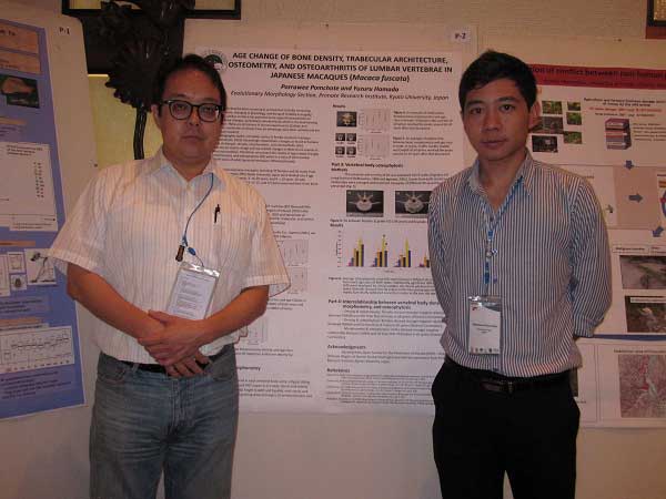
2. To present a paper at the international symposium on "Diversity and Evolution of Asian Primates" at Chulalongkorn University, Bangkok from 27-29th August, 2012.
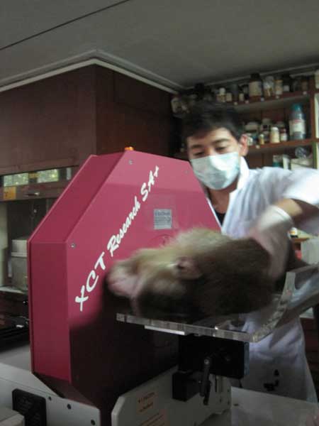
Twenty eight females (aged 19-33 years) and 16 males (aged 21-37 years) of long-tailed macaques (Macaca fascicularis) were chosen for the experiment. The animals were fasted for at least 18 hours before the experiment was conducted. The experiment was performed between 8.00-12.00 h. They were anesthetized by a mixture of ketamine (100 mg/ml) and domitor (10 mg/ml) (10:1) via intramuscular injection. The body weight was recorded, blood sample (5 ml) was drawn from the femoral vein, and serum was separated at 80˚C for bone marker analysis. Bone formation marker, that is, osteocalcin and bone specific alkaline phosphatase, and bone resorption marker, that is, pyridinoline and deoxypyridinoline crosslinks were determined using enzyme-linked immunosorbent assay (ELISA). The animals were placed into the peripheral quantitative computed tomography (pQCT) machine and the bone was determined bone mineral densities (both cortical and trabecular densities) at distal sites of radius bone for 7-10 minutes. The animals were moved and placed on the table and the gross anatomy of lumbar bone was determined using portable X-ray machine for 0.06-0.14 seconds. The bone anatomy was used to indicate the osteoarthritis in animals. During the determination of bone anatomy by the X-ray machine, the researcher and surrounding area were shielded by the X-ray shielding partition. After the inspection was completed, they were injected with antisedan (5 mg/ml) via subcutaneous injection and returned to their cage. They were carefully monitored until they are fully recovered. Polynomial trend line superimposed or linear regression will be used to determine the association between each parameter. The relationship between the three aspects of age-related bone changes; bone mineral density, serum bone marker, and osteoarthritis will be analyzed using partial correlation. Statistical significance will be considered when P value was lower than 0.05.
working
presentation
AS-HOPE Project< > >
|