Anatomical evidence for the involvement of medial cerebellar output from the interpositus nuclei in cognitive functionsXiaofeng Lu, Shigehiro Miyachi, and Masahiko Takada 陸 焼峰, 宮地 重弘, 高田 昌彦 要約:
京都大学霊長類研究所の高田昌彦教授と宮地重弘准教授の研究グループは、米国ミネソタ大学脳科学センターの陸 焼峰准教授との共同研究により、小脳で処理された情報の出口である小脳核(中位核と歯状核)は、運動機能と認知機能に関わる小脳からの信号をそれぞれ部位特異的に仕分けして、大脳や脊髄に出力していることを明らかにした。
内容:
小脳は長年、運動の実行機能を担っていると考えられてきたが、最近では認知機能、特に行動の認知的側面に関わっていることが示唆されている。実際、小脳で処理された情報の出口の一つである小脳核の中位核は連合運動学習などの認知機能に関与していることが知られているが、それを実証する解剖学的知見は得られていなかった。今回、京都大学霊長類研究所の高田昌彦教授と宮地重弘准教授の研究グループは、米国ミネソタ大学脳科学センターの陸 焼峰准教授との共同研究により、小脳核(中位核と歯状核)は、運動機能と認知機能に関わる小脳からの信号を部位特異的に仕分けして、大脳や脊髄に出力していることを明らかにした。研究グループでは、シナプスを越えて神経回路を構成するニューロンをラベルすることができる狂犬病ウイルスを用いて、それぞれ運動機能と認知機能の高次中枢である、大脳皮質の一次運動野や前頭前野(特に46野)に多シナプス性に入力する小脳核ニューロンの分布を解析した。その結果、運動情報は後中位核と歯状核の背側部や前中位核から視床を介して一次運動野に入力するのに対して、認知情報は後中位核と歯状核の腹側部から異なる視床の領域を介して前頭前野に入力することを見出した。このことは、後中位核が歯状核と同様に運動機能と認知機能に関わる2つの出力チャネルを持っているのに対して、前中位核は運動チャネルのみを持っていることを示しており、小脳失調の際に発現する運動障害や認知障害の治療ターゲットを特定するのに寄与できると考えられる。なお、本研究の成果は米国科学アカデミー紀要(2012年109巻46号)に発表された。 Proceedings of National Academy of Science USA; 109(46): 18980-18984, 2012 図1:小脳核から一次運動野および前頭前野への多シナプス性入力様式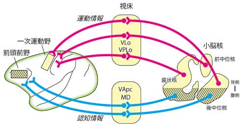
小脳で処理された運動情報が小脳核のうち後中位核と歯状核の背側部や前中位核から視床を介して一次運動野に入力するのに対して、認知情報は後中位核と歯状核の腹側部から前頭前野に入力する。
Although the cerebellar interpositus nuclei are known to be involved in cognitive functions, such as associative motor learning, no anatomical evidence has been available for this issue. Here we used retrograde transneuronal transport of rabies virus to identify neurons in the cerebellar nuclei that project via the thalamus to area 46 of the prefrontal cortex of macaques in comparison with the projections to the primary motor cortex (M1). After rabies injections into area 46, many neurons in the restricted region of the posterior interpositus nucleus (PIN) were labeled disynaptically via the thalamus, whereas no neuron labeling was found in the anterior interpositus nucleus (AIN). The distribution of the labeled neurons was dorsoventrally different from that of PIN neurons labeled from the M1. This defines an anatomical substrate for the contribution of medial cerebellar output to cognitive functions. Like the dentate nucleus, the PIN has dual motor and cognitive channels, whereas the AIN has a motor channel only. NOV/22/2012
Copyright(C) 2012 PRI ( ). ).
|