Segregated pathways carrying frontally-derived top-down signals to visual areas MT and V4 in macaquesTaihei Ninomiya, Hiromasa Sawamura, Ken-ichi Inoue, and Masahiko Takada 二宮太平, 澤村裕正, 井上謙一, 高田昌彦 私たちが普段見ている視野内のさまざまな物体は、その視覚情報の要素ごとに異なる大脳視覚野で処理されていることがわかっている。例えば、頭頂葉のMT野は主に物体の動きや奥行きの情報を処理しており、側頭葉のV4野は主に色や形の情報を処理している。これらの領域は単純に目に映る物体に反応するだけでなく、記憶や注意などの状況に合わせて活動が変化することが知られている。このような高度な脳機能には前頭前野で形成される認知情報が必須であると考えられる。今回、京都大学霊長類研究所の二宮太平特定研究員、高田昌彦教授らの研究グループは、MT野とV4野に伝達されるトップダウン信号が前頭前野の46野腹側部から発信されていることを明らかにした。46野腹側部は特に作業記憶に重要な役割を担っていて、高次視覚野に対して今どこにある、どのような物体に注目すべきかといった情報を提供している可能性がある。さらに、前頭前野からMT野とV4野に伝達される信号が、眼球運動の中枢である前頭眼野や外側頭頂間溝周辺領域の異なる神経細胞によって中継されていることも明らかになった。以上の結果は、動き・奥行きや色・形といった視覚情報に基づく物体認知に前頭前野から発信されるトップダウン信号が本質的な役割を果たしていることを示唆している。このような前頭前野と高次視覚野との相互作用の神経基盤を理解することによって、注意障害など高次脳機能障害の病態解明、さらには治療法の開発に繋がることが期待できる。 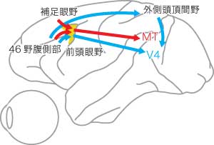
The Journal of Neuroscience, 32(20):6851-6858, 2012 The bottom-up processing of visual information is strongly influenced by top-down signals, at least part of which is thought to be conveyed from the frontal cortex through the frontal eye field (FEF) and the lateral intraparietal area (LIP). Here we investigated the architecture of multisynaptic pathways from the frontal cortex to the middle temporal area (MT) of the dorsal visual stream and visual area 4 (V4) of the ventral visual stream in macaques. In the first series of experiments, the retrograde transsynaptic tracer, rabies virus, was injected into MT or V4. Three days after rabies injections, the second-order (disynaptically-connected) neuron labeling appeared in the ventral part of area 46 (area 46v), along with the first-order (monosynaptically-connected) neuron labeling in FEF and LIP. In the MT-injection case, second-order neurons were also observed in the supplementary eye field (SEF). In the next series of experiments, double injections of two fluorescent dyes, Fast blue and Diamidino yellow, were made into MT and V4 to examine whether the frontal inputs are mediated by distinct or common neuronal populations. Virtually no double-labeled neurons were observed in FEF or LIP, indicating that separate neuronal populations mediate the frontal inputs to MT and V4. The present results define that the multisynaptic frontal input to V4 arises primarily from area 46v, whereas the input to MT arises from not only area 46v but also SEF, through distinct FEF and LIP neurons. Segregated pathways from the frontal cortex possibly carry the functionally diverse top-down signals to each visual stream. MAY/28/2012
Copyright(C) 2012 PRI ( ). ).
|