Reports
Program No.17-003 (Joint Research)
Acquirements of the methods of the dissection in water and the
CT-imaging
By Tomokazu kawashima (Tokyo Women's Medical University)
Period of visit: 26th April 2005 - 24th May 2005.
The main aim of this visit to the Institute for Zoo and Wildlife
Research (IZW) in Berlin, Germany, is to obtain the macroscopic anatomical
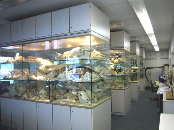
technique for the flesh materials. This method is very convenient for
making a skeleton after a dissection. For obtaining the method, a vocal
tract of a male Saiga was chosen because this field was main theme of my
supervisor, Dr. Frey in IZW.
The dissection was mainly examined using a scissors and forceps.
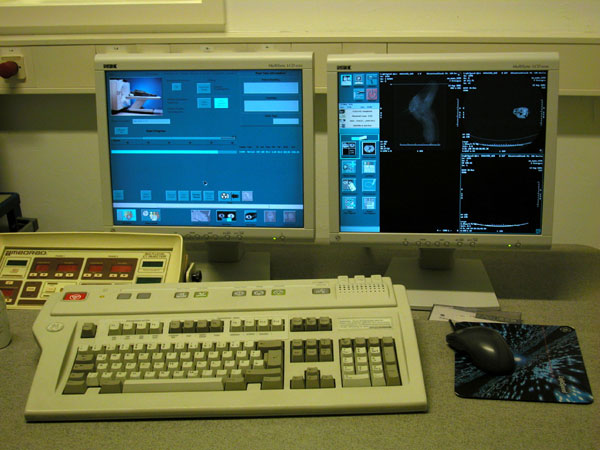
Consecutive dissection steps were documented by digital camera. First, we
carried out a non-destruction analysis of the specimen by mean of CT scan
before a dissection. In this analysis, the material was scanned 1.2mm
thickness by spiral CT and these data were reconstructed. Next, we
dissected the Saiga macroscopically in water using a scissors and
forceps.Consequently, the anatomical differences between the CT-data and
real structure were recognized. In addition, It seems that this method is
not suitable for dissection of the peripheral nervous system.
However, these CT data of the skeleton and the cartilage are
useful as a reference before a dissection.

HOPE Project< > >
|